Tag Archives: Physical Therapy Clinic

Sports Injuries

Sports Injuries and Its Classifications:

INTRODUCTION:

Sports medicine, like all other branches of medicine, aims at the complete physical, mental and spiritual well-being of a sportsperson. A healthy mind in a healthy body is a concept, which is more true to a sportsperson than anybody else is. Positive thinking, fair play and sportsmanship should be the hallmark of a true sportsman. We, the doctors and the therapists, aim to keep a sportsperson physically fit so that the rest of the objectives mentioned above are attained automatically.

Like in other branches of medicine so in sports medicine, prevention is better than cure. To prevent sports injuries, the first step is to ascertain whether a person choosing sports is fit to take it. An unfit person taking up sports is a sure prescription for future sports injuries. A fitness testing for those who wish to take up sports, as their career should include various relevant parameters

However, one has to remember that fitness testing is not done only at the initial stages but needs to be done repeatedly at every stage of an athlete or a sportsperson’s life. The second stage of prevention of sports-related injuries is assessing whether a sportsman is fit enough to resume the sporting activity after the initial layoff. There is nothing more dangerous than an unfit or partially fit person resuming the sporting activity. It may spell a doom to his otherwise flourishing career in sports. A sportsperson has to satisfy certain norms before he can finally be sent back to the field.

CLASSIFICATION OF SPORTS INJURIES

Among the various classifications proposed for sports injuries, the one proposed by Williams (1971) is widely used and recommended.

Williams’ Classification:

Sports Injury Treatment

Among the Consequential Injuries

Primary Extrinsic

  • This is further subdivided into:
  • Human: Black eye due to direct blow.
  • Implemental: May be incidental (as in blow from a hard ball) or due to overuse (blisters from oars).
  • Vehicular: Clavicle fracture due to fall from cycle, etc.
  • Environmental: Injuries in divers.
  • Occupational: Jumper’s knee in athletes, chondromalacia in cyclists, etc

Primary Intrinsic

This could be acute or chronic.

  • Incidental: Strains, sprains, etc.
  • Overuse:
  1. Acute, e.g. acute tenosynovitis of wrist extensors in canoeists.
  2. Chronic, march fracture in soldiers, etc.

Secondary

Short-term: For example, quadriceps weakness.

Long-term: Degenerative arthritis of the hip, knee, ankle, etc.

No Consequential Injuries

These are not related to sports but are due to injuries either at home or elsewhere and are very not connected to any sports (e.g. slip and fall at home).

COMMON SPORTS INJURIES

Sports medicine usually deals with minor orthopedic problems like soft tissue trauma. Very rarely, there may be serious fractures, head injuries or on the field deaths. There is nothing unusual about these injuries except that a sportsperson demands a 100 percent cure and recovery while an ordinary person is satisfied and happy with a 60-80 percent recovery. The difference is because of the desire of the sports person to get back to the sport again, which requires total fitness.

The following are some of the most common sports -related injuries one encounters in clinical practice.

Upper Limbs

  • Shoulder complex
  1. Rotator cuff injuries
  2. Shoulder dislocations
  3. Fracture clavicle
  4. Acromioclavicular injuries
  5. Bicipital tendinitis or rupture.
  •  Elbow
  1. Tennis elbow
  2. Golfer’s elbow
  3. Dislocation of elbow.
  • Wrist
  1. Wrist pain
  2. Carpal tunnel syndrome
  • Hand
  1. Mallet injury
  2. Baseball finger
  3. Jersey thumb
  4. Injuries to the finger joints.

Lower Limb

  • Hip
  1. Iliotibial or tract syndrome
  2. Quadriceps strain
  3. Hip pain
  4. Groin pain due to adductor strain
  • Knee Joint
  1. Jumpers Knee
  2. Chondromalacia
  3. Fracture patella
  4. Knee ligament injuries
  5. Meniscal injuries.
  • Legs
  1. Calf muscle strain
  2. Hamstrings sprain
  3. Stress fracture tibia
  4. Compartmental syndrome of the leg.
  • Ankle Injuries
  1. Ankle sprain
  2. Injuries to Tendo-Achilles
  3. Tenosynovitis.
  •  Foot
  1. March fracture
  2. Jones fracture
  3. Forefoot injuries
  4. Injuries of sesamoid bone of the great toe.
  • Head, Neck, Trunk and Spine
  1. Head injuries
  2. Whiplash injuries
  3. Rib fractures
  4. Trunk muscle strains
  5. Abdomen muscle strain
  6. Low backache

All these injuries have been discussed in relevant sections.

Investigations

These are the same as for any ortbopedic-resared disorders and consist of plain X-ray, CT scan, bone scan, MRI, arthroscopy, arthrography, stress X-rays etc.

TREATMENT OF SPORTS INJURY

This is discussed under three headings prevention, treatment proper and training.

Preventive Measures

The best way to treat a sports injury is to prevent it from happening. Nothing is better than preventing the injury.

Treatment

Treatment of individual sports-related disorders is discussed under suitable sections. However, a mention is made here of the general principles of treatment which is applicable to all sports injuries.

General Principles

  • Concept of RICEMM: This sums up the early treatment methodology of sports injuries and consists of:

R-Rest to the injured limb

I-Ice therapy

C-Compression bandaging

E- Elevation of the injured part

M- Medicines like painkiller s, etc.

M- Modalities like heat, straps, supports, etc.

  • After immobilization and rest, early vigorous exercises should be commenced at the earliest to prevent muscle weakness and atrophy.
  • To prevent joint stiffness, early mobilization ha s to be done first by passive movements and later by active movements. To improve the strength, resistive exercises are added.
  • Unlike the conventional once a day treatment, a sportsperson needs to be seen at least 2-3 times a day.
  • As mentioned earlier, allow resumption of sporting activity only after the sportsperson assumes 100 percent fitness.
  • Mind training is as important as physical training. By repeated counseling, improve the psychological status of the patient to avoid depression, anxiety and negative attitudes, which may develop during the injury.
  • Orthopedic and surgical treatment to be undertaken at appropriate situations.

Training

The physiotherapist has to train a sportsperson in various exercises to enable him to keep his fitness level very high. After conducting a fitness testing, the therapist has to subject an athlete to various forms of exercises to increase the endurance, strength, running, weight bearing, etc. The following are the various forms of exercises.

Measures of Relaxation

After the vigorous workout mentioned above, the sportspersons are taught methods of relaxation and body stretches. Before an athlete or a sportsperson resumes his sporting activities, a fitness testing is carried out and only then, he is allowed to take to the sports provided he is 100 percent fit.

Ankle Fractures

Broken Ankle (Ankle Fracture) – Types, Treatments, Complications

Ankle fractures are the fractures involving the distal end of tibia and fibula. Ankle fractures are common injuries and can vary from a stable fracture to a complex, unstable fracture dislocation.

Mechanism of Injury:

Fractures of the ankle can result from low-or high-energy forces. Fractures due to low-energy forces may be caused by one of the following mechanisms:

1. Rotational stresses to the ankle caused by twisting forces at the ankle joint while walking, running etc. This is the most common mode of injury.

2. Axial stress on the ankle joint results in fracture involving tibial plafond

The high-energy forces, such as road traffic accidents, cause severe injuries, usually fracture dislocations. The pattern of ankle injury depends upon a combination of:

(i) The position of the foot at the time of injury

(ii) The deforming force.

The position of the foot at the time of injury can be supination or pronation and is described first. The deforming force, which can be adduction, abduction, external rotation and vertical loading; is described next. Twisting force produces external rotation. Fall to one side produces adduction or abduction injury. The four most common deforming forces are: supination/external rotation, pronation/external rotation, supination/adduction and pronation/abduction.

Classifications of Ankle Fractures

Lauge-Hansen classified the ankle fractures based on the pathogenesis or the deforming force (i.e. the mechanism of injury). This classification helps in the manipulative reduction of the fracture, if the displacement is understood correctly. The first part of the classification specifies the position of foot during injury and second part of the title specifies the deforming force, for example:

1. Supination-external rotation injury (most common mechanism of injury)

2. Supination-adduction injury

3. Pronation-external rotation injury

4. Pronation-abduction injury

5. Vertical-compression injuries.

However, there is another classification by Danis and Weber which is relatively simple.

Modified Danis-Weber classification: This is based upon the level of fibular fracture and is purely a radiological classification. In this classification, the fibula is considered as the key to the ankle stability. The higher the fibular fracture, the more extensive is the damage to the tibiofibular ligaments and thus greater the instability of the ankle mortise.

Type A:  Fibular fractures below the level of inferior tibiofibular syndesmosis

Fibula: Transverse avulsion fracture at or below the level of ankle joint: or rupture of the lateral ligament complex.

Medial malleolus: Intact or sheared, with almost a vertical fracture.

Posterior malleolus: As a rule intact.

Syndesmosis (Tibiofibular ligament complex): Always intact.

Type BFractures at the level of inferior tibiofibular fibular syndesmosis.

Fibula: Oblique fracture of the fibula at the level of the ankle joint.

Medial malleolus: Avulsion fracture (fracture line horizontal) or rupture of the deltoid ligament.

Posterior malleolus: Either intact or sheared off as a posterior lateral fragment.

Syndesmosis: Usually, intact or partial rupture.

Type CSuprasyndesmotic fibular fractures unstable injury.

Fibula: Shaft fracture anywhere between the syndesmosis and the head of fibula.

Medial malleolus: Avulsion fracture or rupture of the deltoid ligament.

Posterior malleolus: Either intact or pulled off.

Syndesmosis: Always disrupted.

Clinical Features:

The patient typically present s with a twisting injury to the foot following which they com plain of inability to bear weight, pain around the ankle and very often swelling around the ankle. Clinically the stability of the ankle joint must be tested by valgus and varus stress under anesthesia.  Associated injury to the tendons and the neurovascular bundles, which run in close vicinity to the joint, has to be ruled out. The state of the skin must be checked. The skin over the deformed ankle may get unduly stretched, resulting into necrosis, if not reduced immediately.

Radiological Features

Antero posterior, lateral and mortise view must be taken to define the exact fracture pattern.

Management

The ankle fractures must be reduced accurately. Since ankle is a major weight joint, any incongruity of the articular surface, or tilt or disruption of the ankle mortise can lead to early osteoarthritis. The aim of the treatment in ankle fractures therefore is:

1. Anatomical positioning of the talus.

2. To obtain a smooth articular alignment of the ankle mortise.

For management and prognosis, ankle fractures may be grouped into stable and unstable fractures, depending upon the position and the talus, and its instability on light stress. This classification is of importance in treatment and prognosis.

Conservative treatment

Conservative treatment is suggested in treating stable fractures viz. isolated fibular fractures without a medial side injury. These fractures can be treated by below-knee plaster casts for 4-6 weeks followed by graduated weight bearing In unstable fractures with displaced talus closed reduction is achieved by manipulating talus under anesthesia and protecting it with above knee plaster cast for 4-6 weeks.

Open reduction and fixation: This is advocated in unstable injuries and in those injuries where the ankle joint is not properly aligned.

Internal fixation is achieved by

1. Tension band wiring

2. Malleola screws

3. Plate and screw fixation for lateral malleolus.

Complications

Major injuries of the ankle may be associated with the following complications:

1. Non union: Neglected fracture of the medial malleolus may go into nonunion. In old injuries reduction of the fracture and the ankle mortise may be difficult impossible.

2. Stiffness of the ankle.

3. Osteoarthritis: If the fracture has not been treated properly leading to incongruity of the articular surface, early osteoarthritis may set in. The patient has chronic pain and swelling of the ankle necessitating ankle arthrodesis.

Archillies Region – Types, Causes & Treatment

Achilles is a common overuse injury that occurs in people of all fitness levels.  The causes can be variable, but one factor that seems to be consistent with all cases is stress to the gastroc and soleus muscles in the calf region with irritation and loading at the tendon insertion at the heel bone.  This stress may be a result of continued forces placed through the tendon structure from activities ranging from standing, walking, exercise, to recreational activity or sport.

History

The athlete with overuse tendinopathy not ices a gradual development of symptoms and typically complains of pain and morning stiffness after increasing activity level. Pain diminishes with walking about or applying heat (e.g. a hot shower). In most cases, pain diminishes during training, only to recur several hours afterwards.

The onset of pain is usually more sudden in a partial tear of the Achilles tendon. In this uncommon condition, pain may be more disabling in the short term. As the histological abnormality in a partial tear and in overuse tendinopathy are identical. We do not emphasize the distinction other than to suggest that time to recovery may be longer in cases of partial tear. A history of a sudden, severe pain in the Achilles region with marked disability suggests a complete rupture.

Types of Achilles

Midportion Achilles tendinopathy

It is important to distinguish between midportion and insertional Achilles tendinopathy as they differ in their prognosis and response to treatment. We briefly review the pathology of Achilles tendinopathy, list expert opinion of the factors that pre dispose to injury, and summarize the clinical features of the condition. The subsequent section details the treatment of midportion tendinopathy.

Treatment of Midportion Archillies

Archillies tendinopathy Level 2 evidence -based treatments for Achilles tendinopathy include heel-drop exercises, nitric oxide donor therapy (glyceryl trinitrate [GTN] patches), sclerosing injections and micro current therapy (see below), In addition, experienced clinicians begin conservative treatment by identifying and correcting possible etiological factors. This may include relative rest, orthotic treatment (heel lift, change of shoes, corrections of malalignment) and stretching of tight muscles. Whether these ‘commonsense’ interventions contribute to outcome is unlikely to be tested. The sequence of management options may need to vary in special cases such as the elite athlete, the person with acute tendon pain unable to fully bear weight, or the elderly patient who may be unable to complete the heel-drops. As always, the clinician should respond to individual patient needs and modify the sequence appropriately.

Insertional Achilles tendinopathy, retrocalcaneal bursitis and Haglund’s disease

These three ‘diagnoses’ are discussed together as they are intimately related in pathogenesis and clinical presentation.

Relevant anatomy and pathogenesis

The Achilles tendon insertion, the fibro cartilaginous walls of the retrocalcaneal bursa that extend into the tendon and the adjacent calcaneum form an ‘enthesis organ’. The key concept is at this site the tendon insertion, the bursa and the bone are so intimately related that a prominence of the calcaneum will greatly predispose to mechanical irritation of the burs a and the tendon. Also, there is significant strain on the tendon insertion on the posterior aspect of the tendon. This then leads to a change in the nature of those tissues, consistent with the biological process of mechanotransduction.

Treatment

Treatment must consider the enthesis organ as a unit, isolated treatment of insertional Tendinopathy is generally unsuccessful. For example, Alfredson’s pain, full heel-drop protocol (very effective in midportion, tendinopathy) only achieved good clinical results in approximately 30%ofcases of insertional tendinopathy. Patients with more than two years of chronic insertional tendinopathy, sclerosing of local neo vessels with polidocanol cured eight patients at eight -month follow-up.

Other Causes of pain in the Achilles region

Achilles bursitis is generally caused by excessive friction, such as by heel tabs, or by wearing shoes that are too tight or too large. Various types of rather stiff boots (e.g. in skating, cricket bowling) can cause such friction, and the pressure can often be relieved by using a punch to widen the heel of the boot and providing ‘donut’ protection to the area of bursitis as it resolves. Referred pain to this region from the lumbar spine or associated neural structures is unusual and always warrants consideration in challenging cases.

Clinical perspective

Acute tendon rupture is most common among men aged 30- 50 years (mean age, 40 years); it causes sudden severe disability. Overuse Achilles tendon injuries-tendinopathy may arise with increased training volume or intensity but may also arise insidiously. Because the prognosis for midportion Achilles tendinopathy is much better than for insertion tendinopathy, these conditions should be distinguished clinically. Most textbooks suggest that rupturelim its active plantar flexion of the affected leg- but beware, the patient can often plantarflex using an intact plantar is and the long toe flexors. The condition that was previously called ‘Achilles tendinitis’ is not truly an inflammatory condition and, thus, should be referred to as ‘Achilles tendinopathy” pathology that underlies the common tendinopathy.

Whether you treat an Achilles tendon rupture with surgery or use a cast, splint, brace, walking boot, or other device to keep your lower leg from moving (immobilizing your leg), after treatment it’s important to follow the rehabilitation program prescribed by your doctor and physical therapist. This program helps your tendon heal and prevents further injury. http://www.alliancephysicaltherapyva.com/

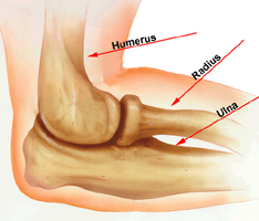
Injuries Around the Elbow

Our elbow joint is made up of bone, cartilage, ligaments and fluid. Muscles and tendons help the elbow joint move. When any of these structures is hurt or diseased, you have elbow problems.Our elbow joint is made up of bone, cartilage, ligaments and fluid. Muscles and tendons help the elbow joint move. When any of these structures is hurt or diseased, you have elbow problems.

Many things can make your elbow hurt. A common cause is tendinitis, an inflammation or injury to the tendons that attach muscle to bone. Tendinitis of the elbow is a sports injury, often from playing tennis or golf. You may also get tendinitis from overuse of the elbow

The injuries around the elbow will be described under the following heads:

  • Fractures of the distal end of the humerus
  • Dislocation of the elbow
  • Fractures of the proximal ends of the radius and ulna.
Fractures of the distal end of humorous:
  • Supracondylar fracture
  • Intercondylar fracture
  • Fracture of the lateral epicondyle
  • Fracture of the medial epicondyle
  • Fracture of the capitellum.

Supracondylar Fracture Of The Humerus

Supracondylar fracture of the humerus is one of the most common fractures in the children, and occurs in the age group of 3-13 years.

Mode of Injury

This fracture is caused by a fall on the outstretched hand.

Displacements

The fracture line runs transversely just above the condyles of the humerus. On the basis of the displacements, fracture is classified into two types:

  • Extension type: In this type the distal fragment is displaced posteriorly.This is the most common type and discussed here.
  • Flexion type: In this rare type, the fragment is displaced anteriorly. Most of the fractures are displaced fractures. In an extension type the distal fragment is:

(i) Displaced posteriorly

(ii) Tilted posteriorly

(iii) Titled medially

(iv) Internally rotated.

 Clinical Features

The child complains of severe pain and swelling in the elbow following a history of fall. The child holds the elbow in a flexed position and resists any movement to the elbow. When brought early, the swelling is less and the following signs can be elicited:

  • There is tenderness over the distal end of humerus,
  • Crepitus can be elicited, although it causes pain and therefore should be avoided,
  • Since the fracture line is above the condyles, and the whole of the distal end of humerus carrying the elbow joint is displaced backwards, normal three bony point relationships is maintained.

When presented late, gross, tense swelling sets in which fills up the hollows around the elbow and obscures the bony landmarks. Sometimes even blisters develop over the elbow. In such a situation the fracture signs cannot be elicited. At the time of injury the distal fragment is displaced posteriorly there by pulling the brachial artery and the median nerve against the sharp distal end of the proximal fragment. This may cause injury to the brachial artery and/or the median nerve. It is therefore important to feel the radial pulse and test the nerve functions at the time of initial examination and make a record of it.

If the distal circulation is affected due to an arterial injury, the following features (5 Ps) may be seen:

  • Pain- severe
  • Pallor
  • Pulselessness
  • Paraesthesia, and
  • Paralysis.

Investigations

Anteroposterior (AP) and lateral view radiographs of the elbow are essential. The AP view shows the fracture line which runs transversely just above the condyles. The distal fragment is displaced and rotated. The lateral view shows the posterior displacement of the distal fragment.

Treatment

An undisplaced fracture is treated above-elbow PoP slab for 3 weeks. A displaced fracture can be treated by one of the following methods:

  • Closed reduction: The fracture is reduced by closed manipulation under general anesthesia. The reduction is obtained by gentle traction to the forearm, manual manipulation of the fragments to align them properly, and then flexing elbow a little beyond 90°. If the radial pulse becomes feeble or disappears during flexion of the elbow, then the elbow is extended gradually till the pulse reappears. The fracture is then immobilized with the elbow in the same position. The fracture may be stabilized by passing.

K-wire percutaneously. The extension type of the fracture is immobilized in an above-elbow PoP slab with the elbow in flexion, whereas the flexion type (less common) of the fracture is immobilized with the elbow in extension. In either case, the plaster is removed after 3 weeks.

  • Traction: The cases which report late (more than one week) with marked swelling and blisters etc. are treated by continuous (Dunlop) traction for 3 weeks.
  • Open reduction: Open reduction of the fracture is indicated when:
  • The closed manipulation fails,
  • The brachial artery is injured and needs exploration, and
  • There is an associated nerve palsy which needs exploration.
  • After open reduction the fracture fragments are fixed internally with Kirschner wires-(commonly called K-wires)

Early complications

These complications occur at the time of injury immediately after.

1. Injury to the brachial artery: This is the most dreaded complication; the brachial artery is injured by the sharp edge of the proximal fragment. The artery may actually be lacerated, thrombosed or may just go into spasm. The blood supply to the flexor muscles of the forearm may be affected resulting into Volkmann’s ischaemia. This requires immediate treatment.

Volkmann’s ischemia: Injury to the brachial artery leads to impairment of circulation to the forearm and hand. There occurs ischemia of the deeper muscles of the flexor compartment of the forearm, such as flexor pollicis longus and flexor digitorum profundus. The muscle ischemia, in turn, leads to compartment syndrome.

Diagnosis:

  • There is severe, sudden increase in pain in the forearm
  • Stretch pain.

There is severe pain in the flexor aspect of the forearm when the fingers are passively extended. This is the most important test and is pathognomonic of muscle ischaemia.

Treatment: The case of Volkmann’s ischaemia must be handled as an absolute emergency because changes may soon become irreversible.

  • Remove tight bandage/splints/plasters etc. immediately.
  • The forearm is elevated and hot bottles are applied to the other three limbs to promote general vasodilation.
  • If no improvement occurs within 2 hours, the operation of fasciotomy is undertaken, if the flexor compartment is tight. In this operation an incision is made from skin down to the deep fascia to decompress the compartment.
  • If the injury to the brachial artery is established by angiography/Doppler, exploration of the brachial artery is undertaken.

2. Injury to the nerves: Median, radial and ulnar nerves may be injured, in that order. In majority of the cases the nerve palsy recovers spontaneously.

Late complications

  • Malunion: Malunion is the most common complication of supracondylar fracture of the humerus and results in a cubitus varus deformity. This deformity occurs if the fracture has been allowed to unite with appreciable medial and internal rotation of the distal fragment.
  • Treatment: If the deformity is unacceptable cosmetically, a corrective osteotomy in the supracondylar area is performed (French osteotomy).
  • Myositis ossificans: Myositis ossificans is ectopic new bone formation around the elbow. This is a common complication which occurs following massage to the elbow after the injury and results in stiffness of the elbow.
Treatment: In the acute painful stage, the elbow is immobilized in an above-elbow plaster slab for about 3 weeks. Otherwise, the main treatment is mobilization of the elbow, despite some pain.

Volkmann’s ischaemia contracture (VIC): Volkmann’s ischaemia, if not treated in time, gradually progresses to Volkmann’s ischaemia contracture.

The ischemic muscles are gradually replaced by fibrous tissue, which contracts and draws the wrist and fingers into flexion. If the peripheral nerves are also damaged by ischaemia, there will be sensory and motor paralysis in the forearm and hand.

Diagnosis: There is marked atrophy of the forearm muscles. There is the characteristic deformity of flexion of the wrist and fingers.

Volkmann’s sign: This sign is characteristic of VIC where the fingers cannot be fully extended passively with the wrist extended: but when the wrist flexed, the fingers can be fully extended passively. This happens because the shortened/contracted flexor muscle-tendon units do not permit full extension of the fingers and wrist simultaneously.

Treatment: In established cases to normal is impossible because irreversible damage has occurred to the import and nerves. However, reconstructive surgery can only improve some function of the hand.

  • Mild cases can be treated by:

(i) Stretching exercises by a physiotherapist and also by the use of

(ii) Turnbuckle splint which gradually stretches the contracted muscles.

  • Moderate cases require a muscle slide surgical operation where the flexor group of the muscles is released from their origin from the medial epicondyle of the humerus and ulna.
  • Severe cases can be treated by shortening of the forearm bones, proximal row carpectomy and wrist arthrodesis etc.

 Considerations

Even after the fracture has healed, full motion of the elbow may not be possible. In most of these cases, the patient cannot fully straighten his or her arm. Typically, loss of a few degrees of straightening will not have an impact on how well the arm will work in the future, including for sports or heavy labor. So treat your problem of Elbow Injury with Physical Therapy. Physical Therapy is the best treatment for the Elbow Injury.

Physical Therapy Treatment for Injuries Around The Ankle Joint


Injuries Around the Ankle Joint…

Ankle sprain is an extremely common complaint with many causes. An ankle sprain occurs when the ligaments surrounding the ankle joint are stretched or torn as the ankle joint and foot is turned, twisted or forced beyond it’s normal range of motion. The most common cause of an ankle pain in athletes is a missed step or a missed landing from a jump or fall. Ankle sprains vary in severity and are classified by the degree of severity.

History

The usual mechanism of injury to the ankle joint is a rotational violence in which the body swings around a trapped foot. Depending on the quantum of force, there may be ligamentous injury or bony injury around the ankle. The exact position of the foot at the time of injury is elicited. Ankle injuries are usually classified by the direction of the force and the position of the foot at the time of injury. Following a tibial plateau fracture or talar fracture, the ankle and subtalar joint may go in for secondary degenerative arthritis, which can present as chronic pain and recurrent effusions of the ankle joint. Presence of knee pain and hip pain should be asked for as the foot and ankle disorders can alter the biomechanics of the limb predisposing the knee and hip to degenerative osteoarthritis.

Examination

Inspection

The foot, ankle and the leg are completely exposed. The position of the foot in relation to the leg is determined. The foot may be displaced anteriorly, posteriorly or sideways depending on the type of injury. The foot is usually displaced laterally in external rotation injuries. It may be displaced medially in adduction injuries and displaced upwards and laterally in vertical compression injuries. In vertical compression injuries with diastasis of inferior tibiofibular joint, the ankle may appear broadened. In fracture dislocation of the talus, the displaced fragment may stretch the skin of the dorsum of the ankle and may impend rupture of the skin.

Palpation

The bony points palpated are:

Lower end of tibia and fibula including the malleoli:

As these bones are subcutaneous, it is easy to find out any fractures, irregularity abnormal mobility. In ligamentous injuries around the ankle, the insertion sites of these ligaments such as anterior talofibular ligament, deltoid ligament may be tender to palpation. To demonstrate the ligamentous injury further, the ankle joint is stressed by giving valgus and varus forces to it. Any abnormal opening out can be demonstrated both clinically and radiologically.

Tarsal bones:

The calcaneum is palpated bidigitally on either side to demonstrate tenderness or thickening or irregularity. In chronic degenerative arthritis of subtalar joint, tenderness and restriction of movements of subtalar joint will be present.

Metatarsal bones:

In Jones fractures the base of the Vth metatarsal is avulsed due to the pull of the peroneus brevis muscle. Fractures of the shaft of the metatarsals are demonstrated by eliciting tenderness on axial pressure over the metatarsal head. Diffuse swelling over the tarsometatarsal joints may be seen in Lanfranc’s fracture dislocation. In ‘march fracture’, there will be diffuse swelling over the neck of lInd metatarsal with pain.

Muscular compartment:

Tendo-Achilles which gets inserted in the calcaneum is frequently injured resulting in loss of active plantar flexion.

Thompson’s test:

Squeezing the calf muscle will cause plantar flexion of the ankle joint. When there is a discontinuity in the tendon, this manoeuvre will not cause plantar flexion.

Movements:

In acute injuries, active movements may not be possible.

Measurement

The leg segment is measured from the medial joint line to the malleolus.The vertical height of the heel is measured from the tip of the medial malleolus to the floor in a standing patient. In fractures of the talus and calcaneum, this height may be decreased. The longitudinal measurement of the foot from the tip of the heel to the tip of great toe and then to the tip of the little toe are measured. Circumferential measurement of the foot at the level of the ankle joint, at the level of maximum arch and at the level of the metatarsal heads are measured and compared with the normal side.

Neurovascular examination

Ankle injuries may rarely be associated with posterior tibial artery and nerve injuries. In Lanfranc’s fracture dislocation, the digital arteries and nerves may get damaged and careful animation is needed to diagnose this. The chronically disabled group usually suffers from the sequelae of old trauma or inflammatory infective or degenerative or neoplastic causes. These patients need to be examined by proper history, detailed examination of the individual bone and joints. After eliciting a detailed history, the examiner should arrive at a provisional differential diagnosis based from the history and then proceed to physical examination. This will help in finding the subtle signs of the disease.

Clinical Features

The Patient typically present with a twisting injury of the foot following which they complain of inability to bear weight, pain around the ankle and very often swelling around the ankle. Clinically the stability of the ankle joint must be tested by valgus and varus stress under anaesthesia, Associated injury to the tendons and the neurovascular bundles, which run in close vicinity to the joint, has to be ruled out. The state of skin must be checked. The skin over the deformed ankle may get unduly stretched, resulting into necrosis, if not reduced immediately.

Physical therapy modalities (such as ultrasound) and manual therapy modalities (such as friction massage) are often used when the acute phase is over.A Physical Therapistis a specialist trained to work with you to restore your activity, strength and motion following an injury or surgery. Physical therapists can teach specific exercises, stretches and techniques and use specialized equipment to address problems that cannot be managed without this specialized physical therapy training.

Are You Suffering from Your Hip Joint..???

Injuries around the hip constitute one of the most difficult injuries to treat and predict the outcome. But the best way to treat your pain is by Physical Therapy without any Burn and Injury. In dashboard injuries, the impact is driven to the knee of the patient which passes on the energy of hip joint causing posterior dislocation of hip.

Examination
Inspection
Attitude: The examination of attitude in a hip joint injury is very useful. In posterior dislocation of hip, the hip will be in flexion, adduction and internal rotation. In intracapsular fracture neck of femur, the lower limb lies in external rotation and minimal shortening. In trochanteric fractures, the lower limb lies in complete external rotation and the limb appears shortened. In anterior dislocation of hip, there will be flexion, abduction and external rotation deformity.

Swelling: In dislocation of hip, the femoral head may be felt either in the gluteal region or in the perineal region or iliac region. In trochanteric fracture, there will be diffuse swelling around the hip and thigh.
Palpation
The bony landmarks to be palpated are:
1.Greater trochanter: The position of greater trochanter helps us in the diagnosis of fractures around the hip. The greater trochanter, anterior superior iliac spine (ASIS) and ischial tuberosity have a constant relationship to each other which will be altered in affections of hip joint and proximal femur. Bryant’s triangle is formed by a line connecting ASIS and greater trochanter, line dropped from the ASIS perpendicular to the floor and the line connecting the greater trochanter and the perpendicular line. The base of the Bryant’s triangle is measured and compared with opposite side. In fractures of the neck and dislocations of hip, the base will decrease to the proximal migration of the trochanter. In posterior dislocations of hip, the greater trochanter will be more anteriorly felt near the ASIS. In anterior dislocations, it will be felt more posteriorly. It should be palpated for tenderness, thickening or irregularity. In subtrochanteric fractures, Bryant’s triangle will not be altered but there will be loss of transmitted movements between the proximal and distal femur.
2.Head of femur: Normally, the femoral arterial pulsation is felt against the head of femur. In dislocations, this resistance is lost thereby altering the intensity of pulsation. The femoral head may be felt posteriorly or anteriorly depending on the type of dislocation. A smooth round bony hard mass which moves with rotational movements of the shaft of femur is nothing but the head of femur. The medial surface of the medial femoral condyle is in the same direction as that of the head of femur. This gives a rough guidance to locate the head in an intact femur.

Neurological examination
In posterior dislocations of hip, the nerve to be commonly affected is the sciatic nerve. The common peroneal part of the sciatic nerve is most often involved than the tibial part manifesting as foot drop.
So Treat your problem of Hip Joint with Physical Therapy at Alliance Physical Therapy which is located in eight prime locations in Northern VA and DC region.http://www.alliancephysicaltherapyva.com/

Treat Spondylolysis and Spondylolisties With Physical Therapy…

SPONDYLOLYSIS AND SPONDYLOLISTIES

These linked conditions generally affect your lower back but may occur in may part of your spine.Spondylolysis occurs when a defect or weakness in a vertebrae develops into a fracture. The vertebra is then at risk of slipping out of line with the vertebrae adjacent to it, leading to spondylolosthesis, which can be debilitating and painful, or may be painless and go unnoticed.

CAUSES
Spondylolysis may start with a minor crack the narrow arch of bone in a vertebra,known as the usually it is the result of a fall or due to strain and overuse .some sports such as cricket and soccer repeatedly put stress on the arches of the vertebrae ,which can lead to minor cracks or breaks.Spondylolisthesis generally develops from spondylolysis ,with the crack widening to a complete break due to further stresses and strains .This break allows the damaged vertebra to slip out of line, which can irritate the linked facet joints and ligaments and possibly trap a nerve.

SYMPTOMS AND DIAGNOSIS
The pain from a displaced vertebra due to spondylolisthesis depends on the degree of slippage. A slight slip may cause little or no pain, while a greater degree of slippage can lead to more intense pain because of the irritation to the spinal joints and ligaments .If your nerve is trapped, there may be some pain numbness, or “pins and needless” in one or both of your legs. Your doctor will make a diagnosis through a physical examination and testing including on X-rayMRI scan and myelogram.

RISKS AND RECOVERY
Back strengthening exercises can help stabilize your posture, but where vertebrae have severely slipped, nerve entrapment can develop that may require surgery. Young people diagnosed with spondylolisthesis should avoid contact sports and activities with a high risk of back injury .A young person who is still growing should be monitored every six months, using X-rays to detect further movements and shift in the spinal column. Once growth stops, the vertebrae are unlikely to slip any farther.

Treat Herniated Disk with Physical Therapy..

Sandwiched between each of the vertebrae in your spinal column is a disk of cartilage that acts as a shock-absorbing pad. These disks have a soft, jellylike center and a tough, fibrous outer layer. A tear in this outer layer will allow some of the soft center to bulge out. This bulge on the nerve roots emerging from the spine in the region of the damaged disk.

CAUSES
Any activity that puts increased pressure on the disks of your spine can lead to a disk hemlation.This can occur in the cervical spine, or, more commonly in the lower back. He general gear and tear that comes with age can also contribute, making middle-aged people susceptible to if they bend suddenly or lift an awkward weight.

SYMPTOMS AND DIAGNOSIS
Depending on the location of the herniated disk, symptoms can vary, but there is usually severe pain and restriction of movement. In the lower back, the pain tends to be a deep unrelenting ache, which may radiate out to your hips; groin buttocks and legs. You may also develop sciatica-a sharp pain, radiating down one leg accompanied by numbness or tingling. Herniated disks can also occur in the neck, causing severe pain that may spread into your shoulders, arms and hands, making it difficult to turn your head or move it backward or forward. You will usually feel pain in only one side of your body. Your doctor will make a diagnosis by performing a physical examination; if your symptoms persist, he may order further tests, such as an MRI or CT scan.

RISK AND DISCOVERY
Recovery from a slipped disk usually takes 4-6 weeks .However if a disk herniated protrudes fully into the spinal curial; it can compress the caudal equine and damage the nerves leading to your legs, bladder and Bowles. This may result weakness and numbness in both legs and the lower part of your body, loss of bladder and bowel control, and even impotence. Although this rarely happens, it is an emergency and you should seek immediate medical help.

Arthritis Types, Symptoms, Causes and Treatment

Literally, many elder people have arthritis, but today it’s not just a problem of the old. Some forms of arthritis affect kids still in diapers, while thousands of people are suffering in the prime of their lives. The general denominator for this condition is joint and musculoskeletal pain, which are grouped together as ‘arthritis.’ Often that pain is a result of swelling of the joint liningArthritis is the most common cause of inability in the USA.

Types of Arthritis:

Arthritis is of two types. One is Osteoarthritis Arthritis and other on is Rheumatoid Arthritis.

Osteoarthritis Arthritis is local or generalized degeneration of the articular cartilage and the formation of “lips and spurs” at the edges of Joints. An exaggeration of the normal aging process.

Rheumatoid Arthritis is an inflammatory disease involving the synovial membranes and the particular structures.

Symptoms:

The main symptoms of Osteoarthritis are:
• Progressive pain
• Joint enlargement
• -lived stiffness in morning
• Difficulty moving
• A grating or crackling sound or sensation in your joints

The main symptoms of Rheumatoid Arthritis are:
• Joint swells with redness and tenderness
• Symmetrical joint involvement is common
• Migrate from joint to joint
• Inflammation around the joints and in other areas

Causes:

Arthritis is cleanly defined as swelling in the joints. There are different types of arthritis, but the two most common types are rheumatoid arthritis and osteoarthritisJoint stiffness and joint pain are the two most common symptoms of arthritis. Those with arthritis may experience more than one inflamed joint. Main Causes are:
• Main Cause of this disease is Inflammation of synovial membrane tissue. This tissue lining the joints in human body and when this tissue becomes swollen, it results to severe pain and stiffness in that body part.
• Being inflexible, unwilling to change, fear, anxiety, depression, deep shock all these are Arthritis Psychological Causes.
• Poor digestion, Hyperacidity, Enzyme deficiency, Poor Skin, Kidney, Gallbladder and Liver activity, spinal imbalance causing reflex conditions as above leading t accumulated toxins which cause an inflammation reaction.
• Excessive use of Meat, soda drinks, coffee, salt, excess refined carbohydrates, sweets, raw vegetable deficiency all these cause arthritis.
• Fatigue can enhance the feeling of pain and more fatigue increase in arthritis pain.

Treatment:

• Raw Vegetable and Juice fasting is the fastest method of attaining result. Fasting period depends upon the patients and conditions and may range from 7-21 days.
• Take Raw Non Citrus Vegetarian diet and avoid coffee, tea, alcohol, sweets etc.
• And one of the best treatment for this is Physical therapy and Hydrotherapy like Hot and cold showers to stimulate general circulation and act as general tonic, Hot compress, Cabinet Bath, Sauna bath, Paraffin bath etc.
• Daily Massage with olive and peanut oil.
• Or sometimes Joint replacement surgery may be required in eroding forms of arthritis.

Best treatment for arthritis is Physical exerciseLow impact aerobic exercise is best. Talk to your medical professional regarding which types of exercises are ideal for you. And people who are suffering from Arthritis due to Physiological cause they must laugh, shed their stress, loose weight, and have more intimacy with outer world. And do regular exercise.

Wrist Pain

Wrist pain is any pain or discomfort that occurs in the wrist. The wrist contains many small bones, cartilage, muscles, blood vessels, and tendons, and is particularly vulnerable to injury. Wrist pain is commonly caused by soreness or injury but may also arise from infectivity or a tumor on the wrist.

Wrist pain is particularly general complaint, and there are many common causes of this problem. It is important to make an accurate opinion of the cause of the symptoms so that suitable action can be directed at the cause.

Causes for wrist pain:

    * Tendonitis

Tendonitis is a standard problem that causes wrist pain and enlargement. This is due to swelling of the ligament cover. Wrist pain treatment which is caused by tendonitis does not need surgical procedure.

    * Sprain

Wrist sprains are regular injuries caused to the ligaments around the wrist joint. Sprains can origin problems by restraining the use of our hands.

    * Carpal Tunnel Syndrome

Carpal tunnel disorder is the state that results from dysfunction of one of the nerves in the wrist. In carpal tunnel syndrome the median nerve is squeezed together or strained off, as it pass through the wrist joint.

    * Arthritis

Arthritis is one of the troubles that can originate wrist pain and complexity in performing daily or general activities. There are a number of causes of arthritis and luckily there are a lot of wrist arthritis treatments.

     * Ganglion Cyst

A ganglion cyst is a type of swelling that frequently occurs over the back of the hand or wrist. These are a sort of fluid-filled capsules. Ganglion cysts are not cancerous. They will not enlarge and they will not spread to other parts of your body.

     * Gout

This occurs when there is too much production of uric acid and a waste product. This forms crystals in joints rather than being excreted in the urine.

    * Pseudogout

This occurs when calcium deposit in the joints (wrists or knees) causing ache and enlargement.

   * Fractures

A wrist fracture is a general orthopedic injury. Patients with a broken wrist may be treated in a cast, or they may need surgical treatment for the fracture.

 When do you need to call your physician about your wrist pain?

If you are not confident about the cause of your wrist pain, or if you do not know the definite cure recommendations for your condition, you should seek medical consideration. Treatments for these situations must be directed at the specific cause of your problem.

Some symptoms seen by a physician include:

  • Inability to carry objects
  • Injury that causes deformity of the joint
  • Wrist pain that occurs at night or while sleeping
  • Wrist pain that persists beyond a few days
  • Failure to flatten the joint
  • Swelling or major bruising around the joint
  • Symptoms of an infection, including fever
  • Any other strange symptoms

What are the best treatments for wrist pain?

 The treatment of wrist pain depends completely on the cause of the problem. Thus, it is very important that you understand the cause of your symptoms before you decide for a treatment plan. If you are uncertain for your diagnosis or for the severity of your condition, you should look for medical guidance before the start of  any treatment.

All treatments listed here are not appropriate for every situation, but may be helpful in your situation.

The first treatment for many common conditions that cause wrist pain is to relax the joint and allow the acute swelling to drop. It is important, however, to use warning when relaxing the joint, because causing no motion to the joint can result a stiff joint. Adjusting your activities so as not to disturb the joint can help prevent worsening of wrist pain.

 Usually Ice and heat pads are commonly used for treatments of wrist pain. But the question arises, which one is the right one to use, ice or heat? And how long should the ice or heat treatments last? Read on for more information about ice and heat treatment or consult your physician.

 Support braces can aid patients who either had a recent wrist sprain injury or those who tend to hurt their wrists easily. These braces act as a tender support to wrist activities. They will not avoid severe injuries, but may help you to carry out simple activities while rehabilitating from a wrist sprain.